

ABOUT ME

Welcome to my blog Artcle slurp. We share latest article for all niche. If you want to publish your article then mail me on articleslurpblog@gmail.com
Jane Hicks

Dental X-rays at the dental office may not rank high on the list of your favorite things, but it shows dental providers a lot. X-rays help dentists to see the condition of your root, teeth, jaw placement, and facial bone composition. They also help local dentist near me to identify and treat dental issues early in their beginning stage. An X-ray is a form of energy that can flow or travel through, or solid objects can absorb it.
Teeth and bones absorb this energy and show up in the X-rays as the light color areas. X-rays pass through less dense objects like cheeks and gums and appear as dark areas on X-ray film. According to a General Dentist Near Me, X-rays can help identify issues they cannot see with an oral exam. Treating and finding programs early in their development may save money, avoid discomfort and pain, and even save your life.
There are two main types of Digital Dental X-Rays: extraoral (when the X-ray film is placed outside the mouth) and intraoral (the X-ray film is outside the mouth). Intraoral X-rays are the most common type. Different intraoral X-ray types exist. Each exhibits other tooth characteristics.
It shows the upper and lower teeth in detail in one mouth area. Each bitewing shows the level of the underlying bone from the tooth’s crown (the exposed surface). The changes in bone density brought on by gum disease can also be seen on bitewing X-rays, which can detect dental decay between the teeth. Bitewing X-rays can also assess the proper fit of various restorations, including dental bridges or crowns surrounding a tooth. Additionally, it can identify or detect any wear or degradation of dental fillings.
From the crown to the region beyond the root where the tooth is attached to the jaw, the entire tooth is visible on Periapical X-Ray. On each periapical X-ray, each tooth in a particular upper or lower jaw region is visible. X-rays at the root’s periapical region check for unanticipated changes in the surrounding bone structures.
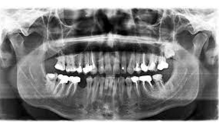
According to family dental care, the entire mouth, including every tooth in the upper and lower jaws, can be seen on a single X-ray. This X-ray can locate fully and partially emerging teeth, locate impacted teeth, and find tumors.
While presenting a specific layer or “slice” of the mouth, they obscure other levels. This X-ray only examines structures that are difficult to view due to obstacles from other nearby structures.
It can send images directly to a computer with dental imaging that uses digital 2-D technology. The images can be saved, printed, or seen on a screen in a couple of seconds. Digital imaging offers several additional advantages over traditional X-rays. For example, a tooth image might be enlarged and improved. Even the slightest alterations that escape notice during an oral examination will be easier for your dentist to spot.
The above-provided details and information tell us about dental X-rays and their types. For more informative facts and updates, please visit urbndental.com.
Welcome to my blog Artcle slurp. We share latest article for all niche. If you want to publish your article then mail me on articleslurpblog@gmail.com